Foundations of care
Labs and acid-base
NCLEX labs chapter: CBC and coagulation, lipids and A1c, specimen-draw traps, critical-value actions, cardiac markers, and primary acid-base interpretation.
ClesialReviewed by Sophia Bennett, RN
Contents12 sections
Lab items ask who gets a phone call now, which precautions match the count, which clotting test belongs to which drug, and whether the ABG is a lung problem or a metabolic one.
Critical values: protect, then notify
A critical value is not “abnormal on the printout.” It is a result that can kill or cripple in a short window if nobody acts. Your job is two steps in order: protect the client first (airway, cardiac monitoring, glucose, bleeding or infection precautions), then notify the provider promptly with the number and the clinical picture. Charting and waiting for rounds is the distractor that looks thorough and fails the stem.
| Result pattern | Bedside protection | Then |
|---|---|---|
| Extreme K+ (high or low) with symptoms or ECG change risk | Cardiac monitor; hold offending infusions as ordered; prepare shift/remove pathway | Notify now |
| Symptomatic hypoglycemia | Give glucose pathway per protocol (oral if safe, IV/glucagon if not) | Notify; recheck glucose |
| Platelets critically low | Bleeding precautions; gentle care; no IM/rectal temps | Notify; watch for occult bleed |
| ANC / WBC critically low | Infection precautions; protect from crowds/sick contacts | Fever = emergency notify |
| Heparin aPTT ~3× control | Hold/adjust per protocol; assess for bleeding | Notify; do not ignore |
| INR markedly above therapeutic band on warfarin | Hold warfarin per protocol; assess bleeding | Notify; anticipate vitamin K/orders |
When several labs are abnormal, stack by minutes-to-harm. Extreme potassium with rhythm risk, symptomatic hypoglycemia, airway failure from hypoventilation, and active bleed with crashing platelets outrank a quietly abnormal BUN. Ion replacement detail lives in Electrolytes. Peak and trough timing for drug levels lives in Vital signs and physical assessment — wrong draw time makes the number meaningless.
Safety
Treat the client, not the printer. A critical number without protection and a phone call is an incomplete answer.
CBC: hemoglobin, hematocrit, WBC, platelets
The complete blood count is the exam’s all-purpose hematology snapshot. Hemoglobin and hematocrit describe oxygen-carrying capacity. White cells and the differential describe infection and marrow stress. Platelets describe clot-forming capacity. Adult reference bands vary by lab and sex; learn direction and hazard, not a fake universal exact number for every stem.
| Panel piece | Adult orientation (typical bands) | Nursing meaning |
|---|---|---|
| Hemoglobin (Hgb) | Often roughly ~12–16 g/dL (female) and ~14–18 g/dL (male); lab-specific | Low = anemia picture: fatigue, pallor, tachycardia, activity intolerance |
| Hematocrit (Hct) | Often about 3× Hgb (rough rule of thumb) | Low tracks with anemia; high can reflect dehydration or polycythemia context |
| WBC | Often about 5,000–10,000/mm³ (lab-specific) | High: infection/inflammation/stress. Very low: infection risk |
| Platelets | Often about 150,000–400,000/mm³ | Low: bleeding risk. Very high: thrombosis context (less common on exams) |
WBC differential cues
The differential tells you which white cell line is reacting. Neutrophils rise with many bacterial infections and acute stress. A “left shift” (more bands/immature neutrophils) means the marrow is pushing young cells into the fight. Lymphocytes often rise with viral patterns. Eosinophils tip toward allergy or parasite contexts on classic stems. You do not diagnose from one percent alone — you match the differential to the fever, wound, and exposure story.
- Neutrophilia + fever + local signs → infection workup, not “watch only.”
- Severe neutropenia (very low ANC): hand hygiene, avoid sick contacts, no fresh flowers/standing water per protocol, cooked foods when ordered.
- Fever in severe neutropenia is an emergency pathway even if the number looks “only” 100.8°F (38.2°C).
Platelets and bleeding precautions
Platelets plug vascular holes. When the count falls into the critically low range (stems often use values near 30,000/microliter or lower), spontaneous bleeding risk climbs. Precautions are mechanical: soft toothbrush, electric razor, no IM injections, no rectal temperatures, fall prevention, gentle BP cuff use, and report petechiae, hematuria, gum ooze, or new headache. Full transfusion-reaction and DIC maps live in Bleeding and transfusion.
Chemistry anchors: electrolytes, glucose, kidney
Chemistry panels on the exam are priority puzzles. Sodium about 135–145 mEq/L — Na 150 is hypernatremia; match the name before you teach fluids. Potassium about 3.5–5.0 mEq/L — extremes are cardiac. Calcium and magnesium shift neuromuscular excitability. This chapter owns the call and precautions; mechanism and food teaching live in Electrolytes.
| Lab | Typical adult band (orientation) | Exam priority |
|---|---|---|
| Sodium | ~135–145 mEq/L | Neuro status; correct slowly when ordered; name the imbalance |
| Potassium | ~3.5–5.0 mEq/L | Cardiac monitor; never IV push K+; notify extremes |
| Glucose (fasting orientation) | Often ~70–99 mg/dL fasting (lab/protocol vary) | Symptomatic hypo first; severe hyper with acidosis → DKA/HHS pathway |
| BUN | Often ~10–20 mg/dL | Rises with renal impairment, GI bleed, dehydration, high protein load |
| Creatinine | Often ~0.6–1.2 mg/dL (sex/muscle mass vary) | Better GFR marker than BUN alone; rising Cr = renal/perfusion concern |
| HbA1c | Often a goal under about 7% for many adults (individualize) | About 2–3 months of average glucose; not a same-hour hypo test |
| Lipids (fasting panel) | LDL and triglycerides up = risk; HDL is the protective fraction | Teaching and statin context; not an ACS rule-out |
Glucose critically low with confusion or diaphoresis is a minutes problem: treat per hypoglycemia protocol, then notify and recheck. Critically high glucose with abdominal pain, Kussmaul breathing, or ketones pushes toward DKA care in Diabetes and glycemic emergencies. BUN and creatinine rising together suggest the kidney is not clearing waste (or perfusion is poor). An isolated BUN bump with a stable creatinine often points more to volume or protein load: still correlate with urine output and the client.
HbA1c answers a different question than a fingerstick. A client can have a reassuring A1c and still be hypoglycemic now, or a high A1c and a normal random glucose after a day of fasting. Use it for control-over-months teaching, not as a substitute for treating diaphoresis and a glucose of 42 mg/dL. Lipid panels on exams are mostly teaching: elevated LDL and triglycerides plus low HDL fit cardiovascular risk and statin counseling in Lipid-lowering therapy. They do not clear chest pain.
Specimen traps: the number is only as honest as the draw
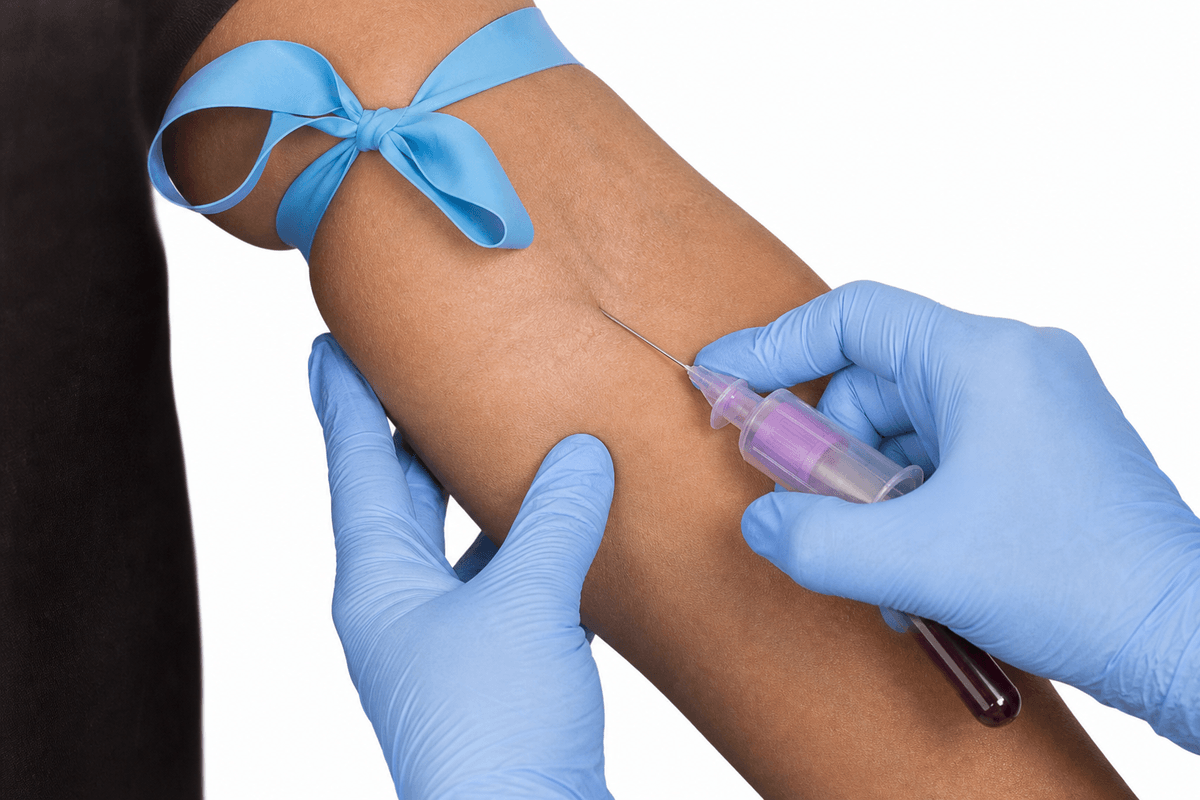
A critical potassium or a wild aPTT can be the client, or it can be the tube. Hemolysis from a traumatic stick dumps intracellular potassium into the sample. A tourniquet left tight for a long wait does the same: cells leak potassium and the printout looks like hyperkalemia. Drawing “labs” from a heparinized central line or from above an infusing heparin drip contaminates coagulation tests and can fake a high aPTT or a distorted potassium. The exam wants a peripheral venipuncture, prompt tourniquet release, and a redraw when the number does not match the client.
| Trap | Why the number lies | Nursing move |
|---|---|---|
| Prolonged tourniquet | Venous stasis and hemolysis raise K+ | Release as soon as flow starts; do not leave it on while you hunt labels |
| Draw from a heparinized line | Heparin contaminates aPTT; residual fluid dilutes or alters electrolytes | Peripheral stick, or a protocol waste/flush sequence the stem actually gives |
| Above an infusing IV | The infusate is in the sample | Other arm, or distal to a stopped peripheral line per policy |
| Hemolyzed chemistry | Cell burst raises K+ (and can alter other ions) | Redraw; do not treat a hemolyzed hyperkalemia as fact if the ECG is calm and the draw was traumatic |
Match the client before you treat the printer. Peaked T waves plus a high K+ is a real emergency whether or not the last stick was pretty. A sky-high K+ with a relaxed ECG after a screaming tourniquet and a hemolyzed comment on the slip is a redraw, not calcium-then-insulin on autopilot. Still protect until you know. Peak and trough timing for drug levels lives in Vital signs and physical assessment.
Coagulation: PT/INR and aPTT
Clotting tests answer two exam questions: is the client protected from clot on therapy, and are they over-anticoagulated. Warfarin (vitamin K antagonist) is monitored with PT/INR. Unfractionated heparin is monitored with aPTT (anti-Xa in some systems: follow the stem). Mixing up the tests is a classic miss.
| Test | Drug it usually tracks | Therapeutic orientation | Too high means |
|---|---|---|---|
| PT / INR | Warfarin | Many goals near INR 2–3; some valves higher per order | Bleeding risk; hold/adjust; assess bleed; vitamin K as ordered |
| aPTT | Unfractionated heparin | Protocol therapeutic band (often ~1.5–2× control) | About 3× control with or without bleed: hold/reduce per protocol; notify |
Heparin aPTT about 3× control means the client is over-anticoagulated relative to the usual therapeutic band. Even without active bleeding, anticipate hold or reduce per protocol and notify — do not “continue and recheck later” as if the number were still in range. The opposite trap is stopping every anticoagulant forever when the real ask is a protocol adjustment and a call. Anticoagulation teaching depth also sits in Vascular disorders and anticoagulation.
Cardiac markers: pattern, not a single stamp
Cardiac troponin rises when myocardial cells are injured. It is the preferred ACS marker on modern stems. CK-MB also rises with cardiac muscle injury but returns toward baseline sooner, so serial sets still matter for timing questions. Myoglobin rises early but is less specific. The nursing move is not to memorize a fake universal cutoff for every lab — it is to escalate chest pain plus rising markers, repeat sets as ordered, and start the ACS pathway while you wait for the next draw.
| Marker | What it tells you | Nursing angle |
|---|---|---|
| Troponin I/T | Myocardial injury; stays up longer | Rising + symptoms → ACS escalation; serial draws |
| CK-MB | Cardiac muscle fraction; shorter window | Supports timing/reinfarction questions with serial enzymes |
| BNP / NT-proBNP (context) | Ventricular wall stretch | Supports heart-failure congestion picture; not a lone ACS rule-out |
A normal first troponin does not clear a client who still has ischemic pain — early draws can miss the rise. Keep monitoring, give ordered ACS care, and expect repeats. Rhythm and STEMI maps live in Acute coronary syndromes and ECG rhythms.
Primary acid-base disorders
Read an ABG in a fixed order. pH first: below the normal band is acidosis; above is alkalosis. Then ask which partner moved with that pH. Carbon dioxide is the respiratory acid — it rises when the lungs are not blowing it off, and it falls when the client is hyperventilating. Bicarbonate is the metabolic base — it falls when acid is added or base is lost, and it rises when acid is lost from the gut or kidneys retain base. That is why respiratory disorders show pH and PaCO2 moving opposite, and metabolic disorders show pH and HCO3 moving the same direction (ROME).
| Disorder | ABG fingerprint | Common cause on exams |
|---|---|---|
| Respiratory acidosis | ↓pH, ↑PaCO2 | Hypoventilation, opioids, COPD |
| Respiratory alkalosis | ↑pH, ↓PaCO2 | Hyperventilation, panic, pain |
| Metabolic acidosis | ↓pH, ↓HCO3 | DKA, diarrhea, renal failure, lactic acidosis |
| Metabolic alkalosis | ↑pH, ↑HCO3 | Vomiting, NG suction, diuretic losses |
Respiratory acidosis is a ventilation failure, not a “lung infection” label by itself. Opioids, oversedation, or severe COPD leave CO2 trapped → PaCO2 up, pH down. First bedside move is support breathing and reverse the driver when ordered — calling it metabolic acidosis because the client “looks sick” is the classic miss. Respiratory alkalosis is the reverse: panic, pain, or acute hyperventilation blows CO2 out → PaCO2 down, pH up; numb lips and tingling are often hypocapnia, not a primary metabolic story.
Metabolic acidosis means the blood lost base or gained non-respiratory acid: DKA ketones, lactic acid from poor perfusion, diarrhea (bicarb leaves in stool), or failed renal acid handling. Metabolic alkalosis is often acid lost from the stomach — prolonged vomiting or NG suction — or diuretic-driven losses that leave relative HCO3 high. Worked pattern check: pH 7.29 with PaCO2 61 after opioids → respiratory acidosis; pH 7.51 with PaCO2 28 in panic → respiratory alkalosis. Fix the breathing problem; do not start by chasing bicarbonate for a pure CO2 story.
Compensation: the other system tries to help
Compensation is the secondary system moving to pull pH toward normal. Lungs adjust CO2 in minutes; kidneys adjust HCO3 over hours to days. If pH is still abnormal and the secondary number has barely budged, call it uncompensated and treat the primary disorder. If pH is closer to normal while both CO2 and HCO3 have moved, call it partial or full compensation per the numbers — still treat the cause, not the compensatory number as the disease.
- Uncompensated: pH abnormal; secondary partner not meaningfully moved yet.
- Partial: pH still off; secondary partner has started to move the helpful direction.
- Fully compensated: pH back in band; both partners abnormal — the primary problem may still be present.
- Panic hyperventilation: coach slow breathing; stay with the client; paper-bag tricks are not the modern default.
- DKA trends: falling glucose/ketones with improving pH/HCO3 means the plan is working.
For panic hyperventilation, coach slower breathing and stay with the client — do not leave numb-lip hyperventilation alone while you argue about paper bags. In DKA, improving glucose and ketones with rising pH/HCO3 means the insulin and fluid plan is working; a worsening acidemia means escalate, not reassure.
Priority map
| Picture | First move |
|---|---|
| Four labs, one critical K+ | Protect rhythm; notify now |
| Platelets 30,000 | Bleeding precautions; notify |
| ANC low + fever 100.8°F | Emergency infection pathway |
| aPTT 3× on heparin | Hold/reduce per protocol; notify |
| K+ 6.8 after a tight tourniquet, ECG normal | Redraw before treating a fake hyperkalemia |
| INR 5.8 on warfarin, gum ooze | Hold warfarin; notify; bleeding care |
| Chest pain + rising troponin | ACS pathway; serial markers |
| pH 7.29, CO2 61 after opioid | Respiratory acidosis; support breathing |
| pH 7.51, CO2 28 in panic | Respiratory alkalosis; slow breathing |
Revision
Must know
- 1Critical values need protect-then-notify: stabilize airway, rhythm, glucose, bleeding, or infection risk first, then call with the number and the clinical picture.
- 2Adult Hgb roughly mid-teens g/dL (sex-specific bands); low Hgb/Hct means less oxygen-carrying capacity. Match anemia care to the picture, not the printout alone.
- 3WBC elevation suggests infection/inflammation stress; severe neutropenia (very low ANC) means infection precautions, and fever is an emergency.
- 4Platelets ~30,000/microliter or lower: bleeding precautions. Critically low counts escalate fall and stick risk.
- 5Electrolyte extremes (especially K+) are call-now. Full ion physiology lives in Electrolytes; here the ask is priority and notification.
- 6Glucose critically high or low: treat the client (symptomatic hypoglycemia first), then notify. Do not chart and wait.
- 7BUN/creatinine rise together in renal impairment or low perfusion; creatinine tracks GFR better than BUN alone.
- 8Warfarin → PT/INR. Heparin → aPTT. Therapeutic INR is usually around 2–3 for many indications (higher for some valves per order). aPTT about 3× control is over-anticoagulated: hold/adjust per protocol.
- 9Do not draw chemistry or coagulation from a heparinized line and call it the client’s potassium or aPTT. Tourniquets left tight too long falsely raise potassium. Peripheral stick, release promptly.
- 10Fasting lipid panel: high LDL and triglycerides are risk teaching; HDL is the protective fraction. HbA1c reflects roughly 2–3 months of glucose; it is not a same-day hypoglycemia test.
- 11Troponin rises with myocardial injury and stays elevated longer than CK-MB. Pattern plus symptoms drives ACS escalation.
- 12ABG first look: pH low = acidosis; pH high = alkalosis. Then see if CO2 or HCO3 matches the direction (ROME).
- 13Respiratory acidosis: high CO2 (hypoventilation, opioids, COPD). Respiratory alkalosis: low CO2 (hyperventilation/panic).
- 14Metabolic acidosis: low HCO3 (DKA, diarrhea, renal failure). Metabolic alkalosis: high HCO3 (vomiting, NG suction, diuretics).
Memory hooks
ROME
Respiratory Opposite (pH and CO2 move opposite). Metabolic Equal (pH and HCO3 move same direction).
Protect, then phone
Critical lab: lock down the hazard at the bedside, then notify with the value and the client picture.
Warfarin is INR; heparin is aPTT
Match the drug to the clotting test before you hold, reverse, or continue.
Tourniquet and heparin lines lie about K+
A tight tourniquet or a heparinized-line draw can fake hyperkalemia or a wild aPTT. Stick a peripheral vein and release the tourniquet promptly.
Opioids hold the CO2 in
RR of 8 after opioids with low pH and high CO2 is respiratory acidosis — support ventilation.
On the exam
How it's tested
Stems ask which lab to call first, what precautions fit platelets 30k, which clotting test matches heparin vs warfarin, why a line-draw potassium is untrustworthy, or which ABG matches panic breathing. Distractors treat critical K+ as routine, restart a unit after hemolysis clues, or call hyperventilation metabolic acidosis.
More in foundations of care
All topics- Adult physical assessmentNCLEX adult assessment chapter: exam order, lung and heart findings to report, perfusion checks, neuro/LOC, pain tools, and abdomen red flags.Read
- Electrolytes: sodium, potassium, calcium, magnesium, phosphateNCLEX electrolytes chapter: Na, K, Ca, Mg, and phosphate. Role, low vs high tables, treatment order, and food plates for diet teaching.Read
- Elimination careNCLEX elimination chapter: incontinence types, retention and oliguria, CAUTI prevention, impaction versus diarrhea, enemas, and stoma color emergencies.Read
- Fire safetyNCLEX fire safety chapter: RACE and PASS, oxygen fire risk, evacuate ambulatory clients first patterns as tested, and never use elevators during a fire.Read