Foundations of care
Vital signs and physical assessment
NCLEX foundations chapter: temperature, pulse, respirations, BP, SpO2, and pain, plus orthostatics, assessment order, and peak/trough timing.
ClesialReviewed by Sophia Bennett, RN
Contents14 sections
Foundations items ask whether you measured the right vital the right way and whether the number matches the client in front of you. This chapter owns temperature, pulse, respirations, blood pressure, SpO2, pain, vital-sign craft, and drug-level timing. Head-to-toe technique, lung and heart findings, and abdomen red flags live in Adult physical assessment. Chest-pain perfusion mapping and focused stroke neuro exams stay in Acute coronary syndromes and Stroke and neuro assessment.
What counts as a vital-sign set
A vital sign is a decision tool, not chart decoration. The usual set is temperature, pulse, respirations, blood pressure, oxygen saturation, and pain. Tachycardia with fever means something different from tachycardia with bleeding. Hypotension with cool clammy skin is perfusion failure until proven otherwise. Always pair the number with symptoms, baseline, and trend. A “normal” BP that is 40 points below the client’s usual is still a finding.
| Vital | Adult starting point | What the exam pushes |
|---|---|---|
| Temperature | Often about 97.0–99.0°F (36.1–37.2°C); fever thresholds follow agency/provider rules (many use ≥100.4°F / 38°C) | Route choice, wait times, fever vs hypothermia |
| Pulse | 60–100 beats/min | Apical rules, pulse deficit, amplitude grade |
| Respirations | 12–20 breaths/min | Count without announcing it; depth and pattern |
| Blood pressure | Normal under 120/80 mm Hg (ACC/AHA adult categories) | Cuff size, site bans, orthostatics, Korotkoff |
| SpO2 | Usually 95–100% | Sensor site, fake lows, when to notify |
| Pain | Client report + scale | Reassess after treatment; nonverbal tools |
Assistive personnel may collect many routine vitals, but interpretation, abnormal response, and deciding how often to recheck stay with the nurse. Document clearly and escalate unexpected values with the clinical picture attached.
When to measure
- On first contact / admission and during physical assessment
- Before and after invasive procedures or surgery
- Before, during, and after meds that change heart rate, BP, breathing, or temperature control
- Before, during, and after transfusion
- When the client’s condition changes or they report feeling “different” or sick
- Before/after interventions that stress the system (for example, first ambulation)
- With fever or known infection on a tighter schedule (often every 2 to 4 hours per protocol)
Temperature
Body temperature reflects heat production and heat loss. It runs lower on waking, often peaks in late afternoon or evening, and shifts with environment, exercise, stress, infection, the menstrual cycle, pregnancy, and age. Infants do not regulate heat as tightly as older children and adults. An inability to obtain any temperature reading is not a documentation annoyance. In the very young or older adult it can signal hypothermia, which is an emergency path.
| Site | Relative to oral | Use / avoid |
|---|---|---|
| Oral | Reference for most comparisons | Wait 15–30 min after hot/cold intake, smoking, or gum; place in a posterior sublingual pocket |
| Rectal | About +1°F (+0.5°C) | When other routes fail or oral is blocked (NGT, wired jaw, mouth breathing). Avoid after rectal surgery, with diarrhea/bleeding risk, and in many cardiac clients |
| Axillary | About −1°F (−0.5°C) | Less accurate; dry axilla, arm tight to chest when other routes are not possible |
| Tympanic | About −1°F (−0.5°C) | Check canal first; infection, wax, or discharge can distort the reading |
| Temporal artery | Follow device instructions | Dry forehead; if diaphoretic, many devices allow a site behind the earlobe |
Rectal technique, when it is the right choice: left side-lying, lubricated probe aimed toward the umbilicus, about 1.5 inches (3.8 cm) in an adult and no more than about 0.5 inch (1.25 cm) in an infant. Fever care treats the cause and comfort. Shivering and heavy chilling raise oxygen demand, so warm lightly while you follow antipyretic orders rather than forcing a cold bath that triggers more shivering.
- Fahrenheit to Celsius: (°F − 32) × 5/9
- Celsius to Fahrenheit: (°C × 9/5) + 32
- Follow the device’s instructions. Electronic probes are not interchangeable by guesswork.
Pulse
A peripheral pulse is a palpable pressure wave. Rate alone is not enough. Note rhythm, strength (amplitude), and equality side to side. Changes after ambulation, bathing, or exercise tell you whether the client tolerated the work. When BP falls or circulating volume falls, the heart rate usually rises as compensation. Pain, fever, stimulants, emotions, and hemorrhage push the rate up; many cardiac depressants and aging slow it.
| Grade | What you feel |
|---|---|
| 4+ | Strong, bounding |
| 3+ | Full, increased |
| 2+ | Normal, easily palpable |
| 1+ | Weak, barely palpable |
| 0 | Absent |
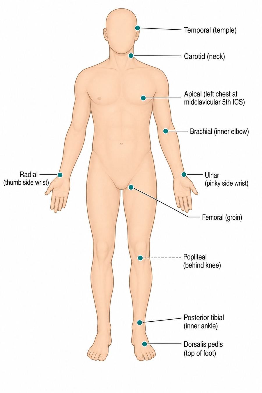
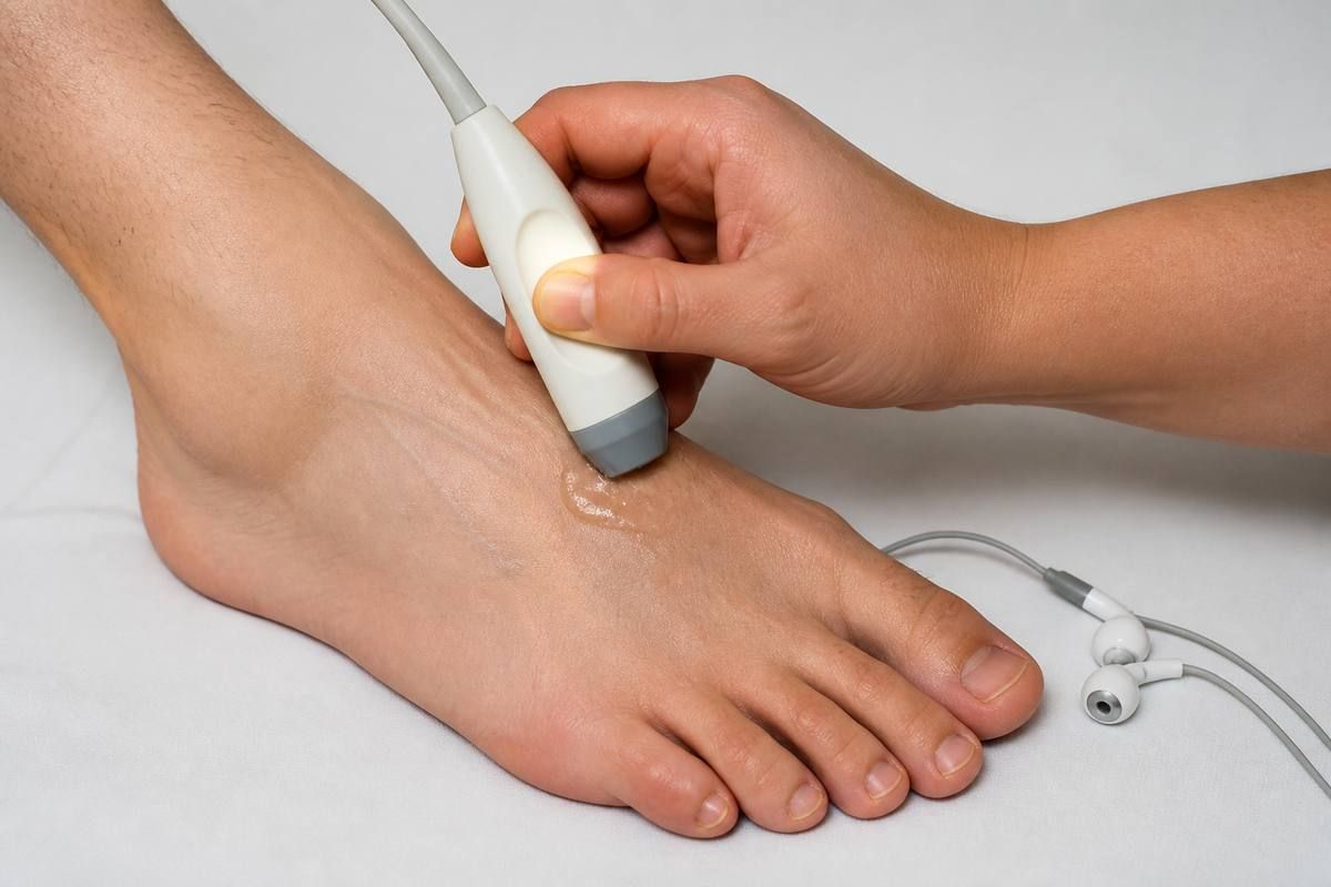
Know the common sites well enough to choose them on sight. Radial at the thumb side of the wrist is the routine count. Apical at the left midclavicular line, fifth intercostal space, is for accuracy. Carotid sits in the groove beside the trachea (never massage both at once). Brachial at the antecubital fossa is for infant counts and BP. Femoral, popliteal, posterior tibial, and dorsalis pedis map limb perfusion. If a pedal pulse is hard to feel, a Doppler ultrasound probe with gel can confirm flow that fingers miss.
Apical count and pulse deficit
Count the apical pulse for a full minute when the radial pulse is irregular, when a heart condition is on the stem, before digoxin or beta blockers as ordered, and in children younger than 2 years. Hold and notify for bradycardia per parameters. A pulse deficit means the apical rate is higher than the radial rate: some ventricular beats are not generating a peripheral wave. One nurse can count apical then radial back to back; two nurses can count simultaneously. Either way, a real deficit is reported.
- Irregular rhythms: count a full minute at the apex when the stem demands accuracy.
- Compare right and left pulses when perfusion or arterial disease is the question.
- Document rate, rhythm, quality, and which site you used.
Respirations
One respiration is one inspiration plus one expiration. Assess rate, depth, pattern, and sounds. Adult baseline is usually 12 to 20 breaths per minute; age changes the expected range. High CO2 or low oxygen drives the rate up. Opioids and other CNS depressants slow it. Head injury or rising intracranial pressure can shallow or slow breathing because the brainstem drive is impaired. Exercise, pain, anxiety, smoking, and body position also move the number.
Count after the radial pulse while you still hold the wrist, or rest your hand lightly on the chest, so the client does not consciously change their breathing. Thirty seconds times two is fine when breathing is regular and the client is stable. Count a full minute when the client is very ill or the pattern is irregular.
Blood pressure
Systolic pressure is the peak during ventricular ejection. Diastolic pressure is the force remaining while the heart rests. Pulse pressure is the difference between them. Adult categories from ACC/AHA are the pattern most exams expect you to recognize, even though treatment thresholds also weigh cardiovascular risk.
| Category | Numbers (adult) |
|---|---|
| Normal | Less than 120/80 mm Hg |
| Elevated | Systolic 120–129 and diastolic less than 80 |
| Stage 1 | Systolic 130–139 or diastolic 80–89 |
| Stage 2 | Systolic at least 140 or diastolic at least 90 |
| Hypertensive crisis | Systolic over 180 and/or diastolic over 120 (with organ-damage cues, escalate as an emergency) |
BP tends to rise with age and stress, runs lower in the early morning, and can read high after smoking or exercise. Have the client rest about 5 minutes, sit with feet flat or lie as protocol says, and stay quiet during the reading. Choose the right site. Avoid an arm with an AV fistula or shunt, the side of mastectomy or axillary node dissection as taught, a traumatized or diseased limb, and an arm with infusing IV fluids when another site exists. The thigh with a popliteal artery is a fallback when both arms are unusable.
- Cuff too small → falsely high BP. Cuff too large → falsely low BP.
- First Korotkoff sound (phase 1) = systolic. Beginning of phase 5 silence = diastolic in most adults.
- If an electronic machine reading looks wrong for the client, confirm with a manual cuff.
- No smoking or exercise in the 30 minutes before a reading when you need accuracy.
Orthostatic (postural) vitals
Orthostatic hypotension is a drop in BP with position change that produces symptoms such as dizziness or near-syncope. Measure BP and pulse supine, then sitting, then standing, waiting the protocol interval (commonly 1 to 3 minutes) after each change. Stop if the client is about to faint. Teaching is slow position change, dangle, and call for help, not “push through the dizziness.” Review antihypertensives and volume status when orthostasis appears.
Pulse oximetry (SpO2)
Pulse oximetry estimates the percent of hemoglobin carrying oxygen. It can warn you about hypoxemia before cyanosis is obvious. A usual adult target is 95% to 100%. Values under about 90% generally need provider notification unless the client already has an established chronic target. Agency parameters win when they are on the stem.
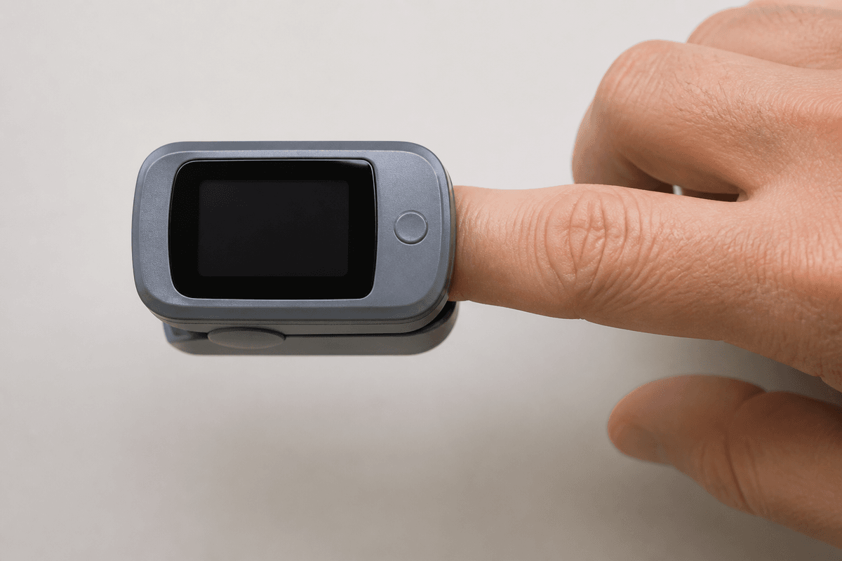
Place the sensor on a vascular pulsatile site: finger, toe, earlobe, nose, or forehead per device. Skip a cold extremity or one with impaired flow. Anything that blocks light transmission can spoil the number: sensor motion, dark nail polish, hypotension, anemia, and peripheral vascular disease. When a reading is low, look at the client first. Support oxygenation as indicated, coach deep breathing when appropriate, and recheck rather than trusting a single odd digit in isolation.
Pain: the sixth piece of the set
Pain is subjective. Ask timing, location, severity, quality, what worsens or relieves it, and what the client already tried. Older adults may show pain as sleep change, gait change, withdrawal, or depression rather than a dramatic report. Clients with dementia or who are unresponsive still hurt. Watch nonverbal cues and use an appropriate behavioral or faces-style scale.
| Pain pattern | Exam angle |
|---|---|
| Acute / transient | Injury, surgery, or acute illness; hours to a few days |
| Chronic persistent | Long-term disorders; months to years |
| Chronic episodic | Breaks of hours to weeks (migraine, sickle cell crisis patterns) |
| Cancer-related | Tumor, procedures, treatment toxicity, or limits on movement |
| Idiopathic | Pain without a clear matching lesion, or out of proportion to findings |
| Nonverbal cues | What to do |
|---|---|
| Grimacing, guarding, rigid posture | Believe the cue; treat and reassess |
| Moaning, crying, irritability, restlessness | Do not wait for a perfect 0–10 score |
| Rising BP, HR, or RR; diaphoresis; nausea | Support ABCs if needed, then pain plan |
| Cannot rate verbally (child, cognitive impairment) | Use Wong-Baker FACES or a FLACC-style tool scored 0–10 |
Wong-Baker FACES pain rating scale
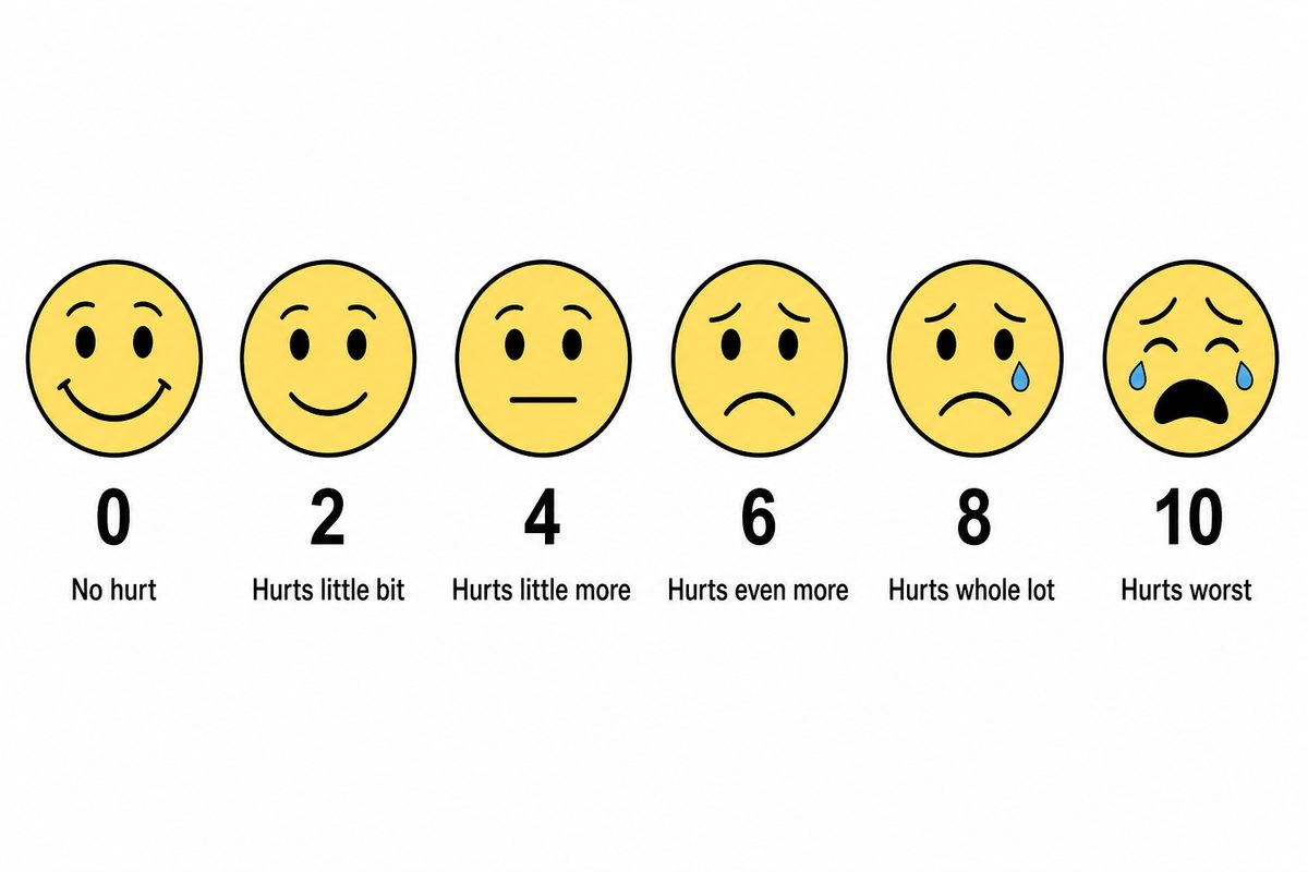
When a child or a client who cannot give a clean 0 to 10 number needs a severity score, offer a faces scale. The client points to the face that matches their pain. Wong-Baker FACES uses even numbers from 0 to 10 so the score still lands on the same 0 to 10 band used for adults. Document the number, not a vague “looked uncomfortable.” FLACC (face, legs, activity, cry, consolability) is the behavioral alternative when the client cannot point.
| Score | Face meaning |
|---|---|
| 0 | No hurt |
| 2 | Hurts a little bit |
| 4 | Hurts a little more |
| 6 | Hurts even more |
| 8 | Hurts a whole lot |
| 10 | Hurts worst |
Reassess after pharmacologic and nonpharmacologic measures. Heat, cold, positioning, binders, and ordered TENS can help selected pains, but crushing chest pain or a client who cannot breathe is not a “finish the pain scale first” moment. Those stems move into cardiac and respiratory chapters once you recognize the emergency.
Vital signs that change the plan
Once technique is solid, the exam asks whether you notice the trend that means bleeding, sepsis, respiratory failure, or drug effect.
| Finding | Priority angle |
|---|---|
| Orthostatic drop + dizziness | Fall risk; slow rises; review antihypertensives/volume |
| Apical bradycardia on digoxin | Hold per parameters; notify |
| Pulse deficit (apical > radial) | Perfusion/dysrhythmia concern; notify |
| New SpO2 drop with distress | Airway/oxygen pathway; do not chase a single sat alone |
| High fever + rigors | Infection workup; comfort; oxygen demand up |
| Post-op falling BP + rising HR | Bleeding/hypovolemia until proven otherwise |
Where technique order lives
Abdominal sequence (listen before you poke), lung and heart findings to report, capillary refill, LOC change, and rigid-abdomen emergencies are taught in Adult physical assessment. Keep that chapter open when the stem is about hands and stethoscopes rather than the vital-sign number itself.
Therapeutic drug monitoring
Some drugs need blood levels because the gap between help and harm is narrow: digoxin, vancomycin, aminoglycosides, and similar agents on exams. A trough is usually drawn immediately before the next dose (lowest level). A peak is drawn after a dose or infusion finishes on a drug-specific clock. If timing is wrong, the lab number cannot guide the next dose safely.
| Level | Typical timing idea |
|---|---|
| Trough | Just before the next scheduled dose |
| Peak | After dose/infusion per protocol (often 30–60 min after IV finishes as tested) |
| Random | Only when ordered, not a substitute for timed levels |
Nursing responsibilities: hold or give as parameters say, draw from the correct line (not from the same lumen that is infusing the drug when that contaminates the sample), label time accurately, and notify for levels outside the ordered band with the clinical picture attached. Lithium and other psych levels also appear in mental-health stems. Same timing discipline.
Priority map
| Situation | First move |
|---|---|
| Dizzy on standing | Orthostatic set; fall precautions; slow rises |
| Digoxin due + HR 48 | Hold per parameters; notify |
| Irregular radial pulse | Apical for 1 full minute |
| Pedal pulse not felt | Doppler before you chart absent |
| SpO2 low + client looks fine | Check sensor/site/perfusion; reassess the client |
| SpO2 low + distress | Support oxygenation and escalate |
| BP needed + AV fistula arm | Use the other arm |
| Vancomycin trough ordered | Draw just before next dose |
| Child cannot rate pain 0–10 | Use an age-appropriate faces or behavioral scale |
| Abdomen / lung / LOC exam question | Open Adult physical assessment |
Revision
Must know
- 1A full set is temperature, pulse, respirations, blood pressure, SpO2, and pain. Pair every number with symptoms, baseline, and trend.
- 2Adult pulse 60 to 100; respirations 12 to 20; SpO2 usually 95% to 100%. Treat agency parameters and the client picture, not a single absolute.
- 3Oral temp after hot/cold drinks, smoking, or gum: wait 15 to 30 minutes. Rectal is usually about 1°F (0.5°C) higher; axillary/tympanic about 1°F (0.5°C) lower than oral.
- 4Do not take rectal temperature after rectal surgery, with diarrhea/bleeding risk, or in many cardiac clients (vagal stimulation).
- 5Apical pulse for 1 full minute when the radial pulse is irregular, before digoxin/beta blockers as ordered, and in children under 2 years.
- 6Pulse deficit: apical rate greater than radial rate means some beats are not reaching the periphery. Notify.
- 7Orthostatic check: BP and pulse lying, then sitting, then standing on the protocol clock (often 1 to 3 minutes after each change). Dizziness plus a drop means slow rises and fall precautions.
- 8Wrong cuff size lies: too small falsely elevates BP; too large falsely lowers it. Avoid fistula arms, mastectomy/lymph-node sides as taught, traumatized limbs, and IV arms when avoidable.
- 9SpO2 below about 90% usually needs prompt notification unless a chronic exception is already established. Nail polish, cold fingers, motion, and poor perfusion can fake a bad or good reading.
- 10Pain is what the client says it is. Reassess after interventions. Wong-Baker FACES (0, 2, 4, 6, 8, 10) or FLACC-style tools fit children and clients who cannot rate 0 to 10 verbally.
- 11Peak/trough: trough usually just before the next dose; peak after infusion per drug protocol. Wrong timing makes the level meaningless.
- 12Head-to-toe technique order, lung/heart findings, cap refill, and rigid-abdomen red flags live in Adult physical assessment.
Memory hooks
Vitals = TPR + BP + sat + pain
Temperature, Pulse, Respirations, Blood pressure, oxygen saturation, and pain. Leaving pain out is an incomplete set on the exam.
Trough = just before
Draw the trough when the drug is at its lowest, usually right before the next dose.
On the exam
How it's tested
Stems ask which route or site is safe (oral vs rectal temp, which arm for BP), how to count apical or catch a pulse deficit, how to run orthostatics, when SpO2 is unreliable, which pain scale fits a nonverbal child, digoxin hold parameters, or when to draw a vancomycin trough. Distractors take BP on a fistula arm, draw a trough hours after the dose, or chase a single number while ignoring bleeding or distress cues. Abdominal exam order and lung/neuro red flags live in Adult physical assessment.
More in foundations of care
All topics- Adult physical assessmentNCLEX adult assessment chapter: exam order, lung and heart findings to report, perfusion checks, neuro/LOC, pain tools, and abdomen red flags.Read
- Electrolytes: sodium, potassium, calcium, magnesium, phosphateNCLEX electrolytes chapter: Na, K, Ca, Mg, and phosphate. Role, low vs high tables, treatment order, and food plates for diet teaching.Read
- Elimination careNCLEX elimination chapter: incontinence types, retention and oliguria, CAUTI prevention, impaction versus diarrhea, enemas, and stoma color emergencies.Read
- Fire safetyNCLEX fire safety chapter: RACE and PASS, oxygen fire risk, evacuate ambulatory clients first patterns as tested, and never use elevators during a fire.Read