Adult health
ECG rhythms: recognize and act
NCLEX ECG map: normal sinus vs AF, VT, VF, and asystole. What the strip means and the first nursing moves.
ClesialReviewed by Sophia Bennett, RN
Contents9 sections
ECG items ask whether you can name the danger and move. Pulse check, CPR, defibrillation, or a calmer rate-control path. MI reperfusion urgency lives in Acute coronary syndromes. Electrolyte-driven peaked T waves live in Electrolytes. This chapter owns common rhythm recognition and the first nurse actions.
How to read a strip under time pressure
Start with the client: responsive? Breathing? Pulse? A perfect label on a dead monitor cable helps no one. Then rate, regularity, P waves, QRS width. Narrow complex usually means the ventricles are being activated over the normal pathway; wide complex raises ventricular origin or aberrancy. And with no pulse, you stop debating labels and run the pulseless algorithm.
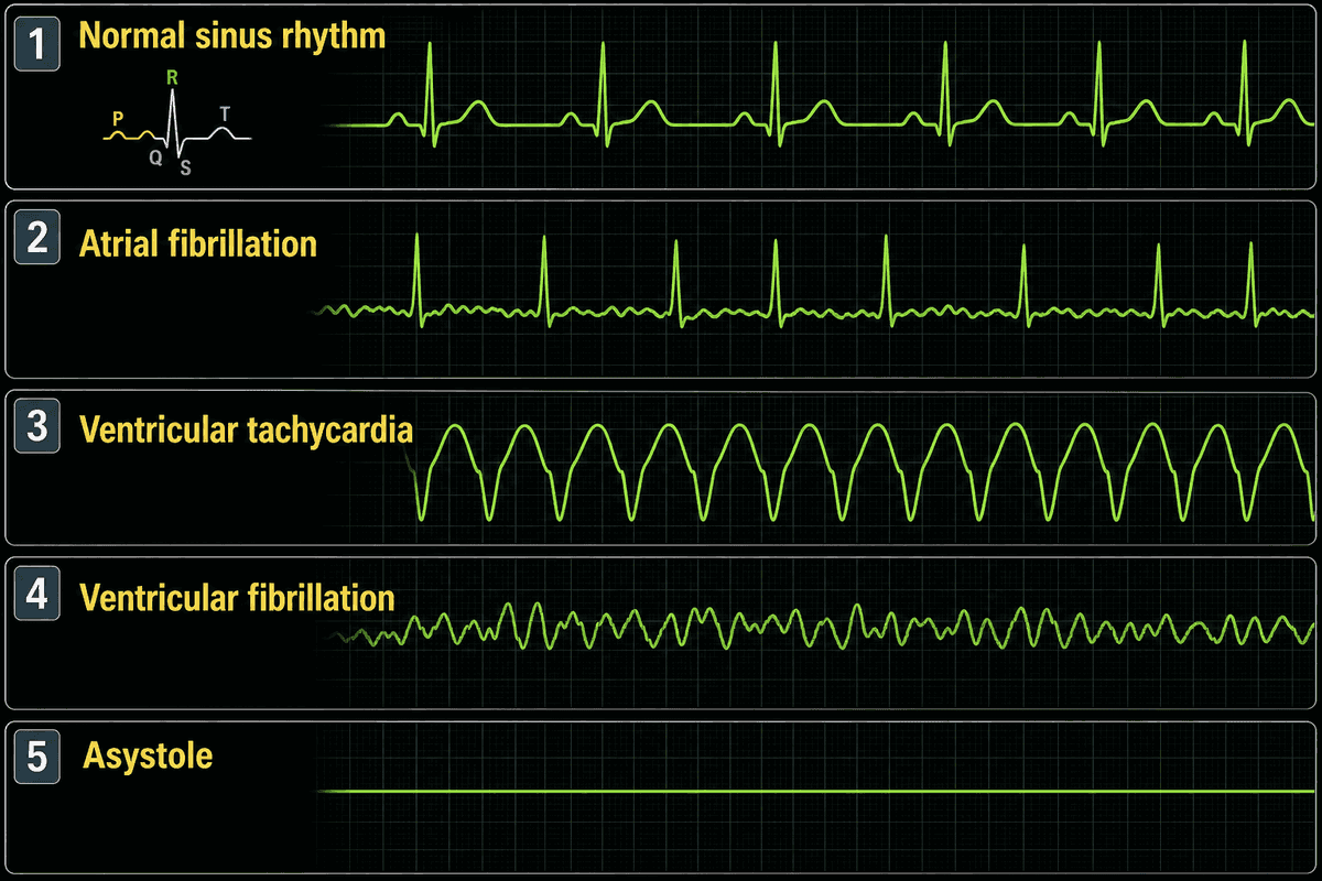
Rhythms you must not confuse
| Rhythm | Strip cues | First nursing lane |
|---|---|---|
| Normal sinus | Regular; P before each QRS; adult rate ~60-100 | Continue assessment; treat the cause of symptoms if any |
| Atrial fibrillation | Irregularly irregular; absent clear P waves | ABCs; rate/symptom control as ordered; embolus risk teaching |
| Ventricular tachycardia | Wide, rapid QRS; may have pulse or not | Pulse check splits the path: pulseless → CPR/defib; unstable with pulse → cardioversion path |
| Ventricular fibrillation | Chaotic, no QRS | Pulseless. CPR and defibrillation; do not wait for a ‘better IV’ |
| Asystole | Flatline (confirm leads) | CPR + epinephrine path; not a defibrillation rhythm |
Why AF matters beyond the squiggle: the atria quiver instead of kick, clots can form, and stroke risk drives anticoagulation conversations. Why VF/pulseless VT matter: cardiac output is effectively zero. Electricity (defibrillation) plus CPR is the chance to restart an organized rhythm. Why asystole is different: there is no fibrillating myocardium to reset with a shock; perfusion comes from compressions and drugs while you reverse causes.
The distractor that looks advanced is synchronizing a shock into VF, shocking asystole, or walking away from AF because ‘the rate looks okay’ while the client is hypotensive and diaphoretic. Edge case: artifact and loose leads mimic VF or asystole. Glance at the client and the electrodes before you announce a code on a brushing-teeth strip.
Safety
Pulseless VT or VF: start CPR and defibrillate as soon as the defibrillator is ready. Do not delay the first shock for routine tasks.
Defibrillation vs synchronized cardioversion
Defibrillation is an unsynchronized shock for pulseless VF or pulseless VT. You are not aiming at a QRS because there is no meaningful QRS to synchronize to. Synchronized cardioversion times the shock to the QRS for selected unstable tachycardias with a pulse (shock, severe hypotension, acute pulmonary edema, ongoing ischemia) so you do not hit the T wave and create VF. Same machine family, different mode and different clinical gate.
- No pulse + VF/VT → defibrillation + CPR.
- Pulse + unstable tachyarrhythmia → synchronized cardioversion as ordered.
- Stable with pulse → ABCs, oxygen as needed, expert consultation / ordered meds. Not random shocking.
Antidysrhythmic drug details (amiodarone, adenosine timing, digoxin hold parameters) appear on stems inside ordered pathways. Know the rhythm lane first so you do not give a rate-control answer to a pulseless client.
Bradycardia, SVT, and heart block
Adult bradycardia under about 50 with poor perfusion (hypotension, altered mentation, shock, ischemic pain) is a treat-the-cause and atropine-as-ordered pathway while you prepare pacing if the client stays unstable. Asymptomatic sinus brady in an athlete is not the same stem. Premature ventricular complexes that are frequent, multifocal, or landing on the T wave raise ischemia and electrolyte questions. Fix hypoxia and potassium before you celebrate a clean strip.
| Pattern | Nursing lane |
|---|---|
| Symptomatic bradycardia | ABCs, atropine as ordered, prepare transcutaneous pacing if unstable |
| Stable SVT (narrow, regular, fast) | Vagal maneuvers then adenosine as ordered; continuous monitor |
| Unstable SVT with pulse | Synchronized cardioversion path |
| High-grade / complete heart block with poor perfusion | Pacing readiness; do not treat as “just slow sinus” |
| Frequent PVCs with ischemia cues | Oxygen, electrolytes, notify; treat the driver |
Adenosine briefly resets the AV node. Warn the client about a transient flush or pause feeling, keep the crash cart mindset, and use a rapid flush technique as taught so the short-acting drug reaches the heart. Heart block that drops output is an electrical delivery problem: atropine may help some nodal blocks, but complete heart block with instability belongs on a pacing path. The distractor shocks asystole, gives adenosine to a pulseless wide-complex client, or walks away from symptomatic bradycardia because “the strip looks organized.”
Pacemakers: spikes, capture, and the generator
A pacemaker writes a spike on the strip, then the chamber it is supposed to depolarize. Atrial pacing: spike then P. Ventricular pacing: spike then a wide QRS. Dual-chamber devices can do both. Capture means the spike actually produces that beat. Sensing means the device sees the client’s own beats and holds fire. Failure to capture is spikes with no QRS (or no P) while the client is still slow and poorly perfused: check connections, atropine and transcutaneous pacing as ordered, notify. Failure to sense paces regardless of native beats and can land a spike on a T wave (R-on-T, VF risk). Do not place defibrillator or cardioversion pads directly on top of the generator can; use an alternate pad map so current does not arc through the device.
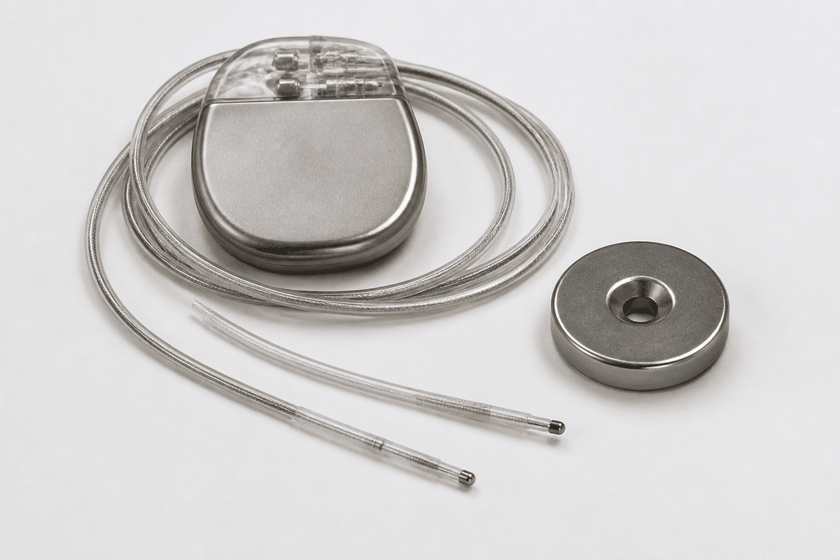
| Finding | Meaning | Nursing move |
|---|---|---|
| Spike then QRS (ventricular) | Capture working | Document; still treat the client |
| Spikes, no QRS, hypotensive | Failure to capture | ABCs, connections, atropine/TCP as ordered |
| Pacing on top of native T waves | Undersensing | Notify; magnet/emergency protocols only as ordered |
| Hiccups or twitching with each spike | Lead stimulation of diaphragm/muscle | Notify; do not ignore |
Client teaching after implant: keep the incision dry as taught, report fever or drainage, avoid heavy ipsilateral arm motion for the prescribed window, and carry identification. MRI compatibility is device-specific; do not send every pacemaker client through MRI on habit. Transcutaneous pacing on an unstable complete block is a now intervention, not a wait-for-the-EP-lab courtesy. The distractor that looks technical is chasing millivolt settings while the client has no pulse, or shocking asystole because you saw pacing spikes on a dead battery strip.
Priority map
| Picture | First move |
|---|---|
| Chaotic strip, no pulse | CPR + defibrillation |
| Wide complex, no pulse | Treat as pulseless VT. CPR/defib |
| Flatline | Confirm leads; CPR + epinephrine path |
| Irregularly irregular, stable | Assess, ordered rate plan, embolus risk |
| Unstable tachy with pulse | Synchronized cardioversion path |
| HR 36 + hypotensive + confused | Symptomatic brady pathway; atropine/pacing as ordered |
| Stable SVT | Vagal then adenosine as ordered |
| Pacing spikes, no QRS, hypotensive | Failure to capture; ABCs and TCP path |
Revision
Must know
- 1Treat the client, not only the strip. Check responsiveness, pulse, and ABCs with every lethal-looking rhythm.
- 2Normal sinus: regular, P before every QRS, rate ~60-100 in adults.
- 3Atrial fibrillation: irregularly irregular, no clear P waves. Rate control, embolus risk, anticoagulation teaching as ordered.
- 4Pulseless VT or VF: defibrillation / ACLS path. CPR until the defibrillator is ready; do not delay for IV starts that block the shock.
- 5Asystole: not a shockable rhythm. High-quality CPR and epinephrine per ACLS; confirm leads/power so you are not treating a disconnected cable.
- 6Unstable tachycardia with pulse (shock, severe hypotension, acute heart failure, ischemia): synchronized cardioversion path as ordered. Different from defibrillation for pulseless VF/VT.
- 7Symptomatic bradycardia: atropine as ordered while you support ABCs; prepare pacing if the client stays unstable.
- 8SVT with a pulse that is stable: vagal maneuvers then adenosine as ordered. Unstable SVT follows cardioversion, not a leisurely teaching moment.
- 9Heart block with poor perfusion is a pacing conversation. Do not treat complete heart block like simple sinus bradycardia with oral teaching alone.
- 10Pacemaker: expect a spike before the chamber it paces. Failure to capture means spikes without QRS (or P) and a slow client: atropine/pacing path, check connections, notify. Failure to sense means pacing on T waves (R-on-T risk). Do not place defibrillator pads directly over the generator.
Memory hooks
Pulse before the pad fantasy
A wide-complex strip without a pulse is a defibrillation/CPR problem, not a ‘watch and give oral meds’ problem.
Irregularly irregular = think AF
No organized P waves and chaotic timing point to atrial fibrillation until proven otherwise.
Flatline is CPR, not shock
Asystole does not get defibrillation; confirm the rhythm and run the CPR/drug path.
On the exam
How it's tested
Stems show a strip description or label and ask the priority action, contrast cardioversion with defibrillation, or show pacemaker spikes without capture. Distractors shock asystole, delay CPR for a second IV, treat pulseless VT like stable sinus tach, or place pads over the generator can.
More in adult health
All topics- Acute coronary syndromesNCLEX ACS chapter: STEMI reperfusion, nitroglycerin and cath-site priorities, cardiogenic shock, tamponade and endocarditis cues, and valvular stenosis vs regurgitation.Read
- Acute respiratory emergenciesNCLEX airway map: failure cues, PE vs pneumothorax vs obstruction, oxygen and positioning priorities, and when to escalate.Read
- AnemiasNCLEX hematology chapter: iron, B12, and folate deficiency teaching, anemia activity tolerance, and sickle-cell crisis priorities with emergency red flags.Read
- Antimicrobial therapyNCLEX antimicrobial chapter: allergy checks, finishing the course, vancomycin flushing, aminoglycoside toxicity, TB drug teaching, and common topical agents.Read