Adult health
Acute respiratory emergencies
NCLEX airway map: failure cues, PE vs pneumothorax vs obstruction, oxygen and positioning priorities, and when to escalate.
ClesialReviewed by Sophia Bennett, RN
Contents8 sections
Respiratory emergencies are ABC items. Protect the airway and gas exchange first, then match the syndrome to the one move that buys time.
Failure cues that raise priority
Gas exchange fails in two main ways that exams mix on purpose. Hypoxemic failure is oxygen delivery collapsing: falling SpO₂, cyanosis, air hunger. Ventilatory failure is CO₂ clearance collapsing: rising PaCO₂ with sleepiness or confusion. Accessory-muscle use and exhaustion mean the client is spending more work than they can sustain; when that tank empties, arrest is next. Stridor or a silent upper airway is not “breathing trouble” in the lung fields. It is an airway that may close.
The distractor is treating a lone low SpO₂ with a casual cannula bump and walking away, or chasing a full history while the client is tiring. Stay, support oxygen or ventilation as ordered, get help when ventilation or the airway itself is failing, and reassess after every change.
| Cue | Why it matters | First instinct |
|---|---|---|
| SpO₂ falling / cyanosis | Hypoxemic failure | Oxygen as ordered; upright if safe; escalate |
| Rising PaCO₂ / somnolence | Ventilatory failure | Support ventilation; call for help |
| Use of accessory muscles / exhaustion | Impending arrest | Do not leave; prepare advanced support |
| Stridor / silent obstruction | Upper airway threat | Airway intervention per protocol |
High-yield syndromes
Once you know gas exchange is at risk, the stem usually hands you one of four acute patterns. Match the onset and laterality, then pick the move that protects breathing while the definitive path starts, not the comfort task that feels caring but wastes minutes.
Pulmonary embolism blocks pulmonary arterial flow, so the classic picture is sudden dyspnea and hypoxia with little or no crackles to explain it; surgery, immobility, and DVT risk tip the odds. Oxygen and rapid notify come before a leisurely teaching session. Pneumothorax (and tension) collapses the lung on one side: unilateral absent or sharply decreased breath sounds and chest pain; tension adds mediastinal shift and obstructive shock, so tracheal deviation with instability is an emergency decompression path, not a “reassess in an hour” finding.
Severe bronchospasm starts loud (wheeze, prolonged expiration) and becomes dangerous when air movement dies: silent chest plus exhaustion means less flow, not improvement. Give ordered bronchodilators/steroids and escalate; chronic rescue-versus-controller order and spacer steps live in Respiratory chronic disease, not here. Aspiration / obstruction is mechanical: if the client cannot speak or move air, run the choking algorithm; if air is moving, support cough and watch closely, and keep NPO when aspiration risk is high.
| Pattern | Classic cues | Do not miss |
|---|---|---|
| Pulmonary embolism | Sudden dyspnea, pleuritic pain, hypoxia; risk: DVT, immobility, surgery | Oxygen + rapid notify; anticipate anticoag / further testing |
| Pneumothorax / tension | Unilateral ↓ breath sounds, chest pain; tension adds tracheal shift + shock | Emergency provider path; high Fowler if ordered; no delay for nonessentials |
| Severe asthma / bronchospasm | Wheeze, prolonged expiration; late: silent chest, exhaustion | Bronchodilators / steroids per orders; escalate silent chest |
| Aspiration / obstruction | Cough, cyanosis, inability to speak if complete | Choking algorithm; NPO when aspiration risk high |
Safety
Tracheal deviation with instability is a now problem. Stay with the client, support oxygen, and get emergency help; do not prioritize a full set of “nice to know” questions.
Oxygen and positioning (exam habits)
Upright / high Fowler uses gravity to drop the diaphragm and open dependent lung. That is why it is the default for dyspnea when the spine and other injuries allow it. Oxygen treats hypoxemia; it does not fix a closed airway or a tiring ventilator pump by itself. When the client cannot protect the airway, suction and readiness for an advanced airway outrank oral teaching every time.
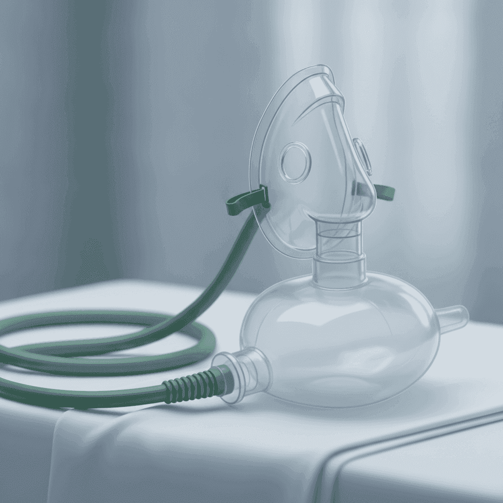
Hypoxic clients still get oxygen on the exam, including many with COPD history. The myth to reject is “never give O₂.” Device choice (cannula → Venturi → non-rebreather, chest tubes) lives in Chest tubes and oxygen devices. Controlled COPD targets and inhaler technique are owned by Respiratory chronic disease; this chapter’s rule is simpler: do not leave hypoxia untreated while you debate the diagnosis.
- Upright / high Fowler for most dyspneic clients unless spinal or other contraindications.
- Titrate oxygen to ordered targets; COPD stems still get oxygen when hypoxic; avoid the myth of “never give O₂.”
- If the client cannot protect the airway, suction and advanced airway readiness beat oral teaching.
Flail chest, ARDS, and status asthmaticus
Flail chest is a free-floating chest-wall segment from multiple rib fractures. Paradoxical motion (segment in on inspiration) plus pain and hypoxia mean oxygen, pain control so the client can breathe, and escalation for possible ventilation. Stabilization follows protocol. Do not strap the chest so tightly that ventilation dies.
ARDS is non-cardiogenic permeability edema: bilateral infiltrates, severe dyspnea, and SpO2 that stays low despite high FiO2. Treat as critical oxygenation failure with ordered PEEP/ventilation strategies. The distractor keeps bumping a nasal cannula and calling it a plan. Status asthmaticus is severe bronchospasm that does not break with initial rescue therapy. Silent chest, exhaustion, and rising CO2 mean intubation readiness, continuous bronchodilators as ordered, and steroids. Chronic inhaler order lives in Respiratory chronic disease.
| Emergency | Do not miss |
|---|---|
| Flail chest | Oxygen, pain control, watch for respiratory failure |
| ARDS | Refractory hypoxemia; critical-care pathway |
| Status asthmaticus | Escalate when rescue fails or chest goes quiet |
| PE with shock | Oxygen + rapid response; anticoagulation/thrombolysis as ordered |
D-dimer and CT angiography belong to the workup when the stem names them. Nursing still oxygenates and escalates first. Immobility, surgery, cancer, and estrogen are PE risk cues that make sudden hypoxia more than “anxiety.”
How to reason under time pressure
Under the clock, force a four-step filter before you fall in love with a detailed distractor. Open airway and moving air first. Failing oxygenation or ventilation next. If either is crashing, support and escalate before you finish the history. Only then sort sudden unilateral chest findings (pneumothorax path) from embolic suddenness without focal lung findings (PE path). Exhaustion, silent chest, or tracheal deviation ends the debate: get help now.
- Is the airway open and is air moving?
- Is oxygenation/ventilation failing right now?
- Match sudden unilateral or embolic cues to PE vs pneumothorax.
- Escalate exhaustion, silent chest, or tracheal deviation immediately.
Revision
Must know
- 1Airway and breathing beat almost every other task when the client is failing gas exchange.
- 2Rising CO₂ + falling LOC = ventilatory failure risk: support breathing, do not just raise the nasal cannula and walk away.
- 3PE pattern: sudden dyspnea, chest pain, hypoxia: oxygen, rapid response / provider, anticipate anticoagulation or further workup.
- 4Tension pneumothorax: tracheal deviation, unilateral absent breath sounds, hypotension: emergency decompression path; do not delay for comfort measures alone.
- 5Complete airway obstruction: intervene for choking; partial with good air movement: encourage cough and monitor.
- 6Position for dyspnea: upright / high Fowler when not contraindicated; prepare for advanced airway if exhaustion or silent chest appears.
Memory hooks
Oxygen first, then the cause
Stabilize oxygenation and ventilation, then chase PE, pneumothorax, aspiration, or asthma path with the stem cues.
Silent chest is not quiet progress
In severe asthma/bronchospasm, loss of wheeze with exhaustion means airflow is collapsing: escalate, do not celebrate “less wheeze.”
On the exam
How it's tested
Stems show sudden dyspnea, unilateral chest findings, or a choking client and ask for the first action. Distractors start detailed teaching, leave a hypoxic client to get a full history, or treat tracheal deviation as a routine assessment finding.
More in adult health
All topics- Antimicrobial therapyNCLEX antimicrobial chapter: allergy checks, finishing the course, vancomycin flushing, aminoglycoside toxicity, TB drug teaching, and common topical agents.Read
- Autoimmune disordersNCLEX autoimmune chapter: SLE flare and nephritis cues, RA versus OA joint care, myasthenia versus cholinergic crisis, and immunosuppression infection risk.Read
- Bleeding and transfusionNCLEX bleeding chapter: transfusion reaction steps, TACO versus TRALI, DIC cues, thrombocytopenia precautions, and clotting failure priorities.Read
- Burns and inhalation injuryNCLEX burn chapter: airway-first inhalation injury, Rule of Nines and Parkland fluid math, burn depth, and early wound priorities.Read