Adult health
Eye and ear care
NCLEX eye and ear chapter: acute glaucoma and retinal detachment emergencies, cataract discharge, drop and ointment technique, timolol caution, and age-based ear drops.
ClesialReviewed by Sophia Bennett, RN
Contents8 sections
Eye and ear items ask whether you can spot the sight-threatening emergencies, put the drop in the right place, and match ear-drop pinna direction to the child’s age.
Emergencies and common eye disorders
Sight-threatening eye stems are pattern recognition, not ophthalmology trivia. Ask: is pressure trapping behind a closed angle, is the retina peeling off, or is this a chronic drop-adherence problem? The wrong first move is often a “helpful” drop or a wait-and-see that costs vision.
| Picture | Think | Move |
|---|---|---|
| Severe eye pain, halos, red eye, nausea | Acute angle-closure | Emergency ophthalmology; no mydriatics |
| Flashes, floaters, curtain shadow | Retinal detachment | Urgent referral; protect the eye |
| Central dark/blurry, edges clearer | Macular degeneration pattern | Ophthalmology follow-up / teaching |
| Painless IOP rise, lifelong drops | Open-angle glaucoma | Adherence; punctal occlusion as taught |
Acute angle-closure is a drainage emergency. The iris crowds the angle so aqueous humor cannot leave → intraocular pressure spikes fast. Halos come from corneal edema; nausea rides the vagal hit from pain and pressure. This is escalate-now ophthalmology. Dilating drops (mydriatics) shove the iris farther forward and worsen the block. The distractor that looks like “treat the red eye” and destroys remaining vision.
Retinal detachment is mechanical separation of retina from its blood supply. Vitreous traction sparks flashes; pigment or blood showers floaters; a curtain or shadow is the field going dark as the peel spreads. Protect the eye, keep the client calm, and get urgent retinal care. Do not wait for “tomorrow’s clinic” or treat it as migraine aura because the headache is mild.
Macular degeneration eats central detail first; peripheral vision often stays usable. Teach safety, lighting, and follow-up. It is not the same sudden curtain story. Open-angle glaucoma is the opposite tempo: painless, slow optic-nerve damage from elevated IOP. Clients stop drops when vision “feels fine,” which is exactly when silent field loss continues. Lifelong adherence and correct technique are the nursing win, not a one-time rescue.
After cataract surgery the wound and the globe hate sudden IOP jumps. Heavy lifting, bending with the head down, straining for stool, and rubbing the eye all push pressure up. Mild scratchiness or tearing can be expected; crushing pain, sudden vision loss, or escalating redness is infection or pressure until proven otherwise. Call, do not coach “wait it out.”
- Cataract postop: avoid IOP spikes (lifting, bending, straining, rubbing). Report severe pain or sudden vision change.
- Sleep with shield if ordered. Mild scratchiness can be expected; crushing pain is not.
Ophthalmic drugs and technique
Eye meds fail in two ways on exams: the drop never reaches the conjunctival sac, or a topical beta-blocker still hits the lungs and heart through the nose. Technique is the intervention. Not just “give the glaucoma drop.”
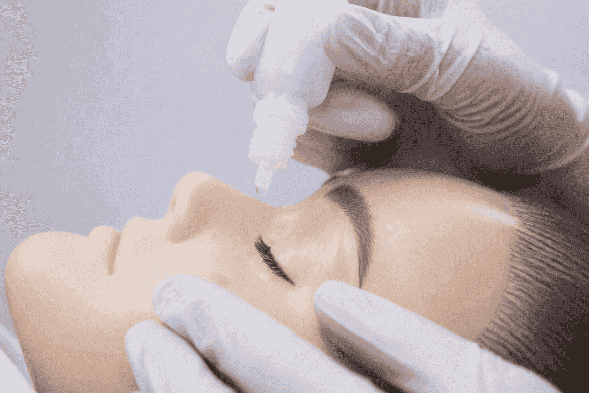
- Wash hands; tilt head back; pull down the lower lid to make a conjunctival sac.
- Drop into the sac. Tip does not touch the eye.
- Close gently; press the inner canthus (punctal occlusion) for about a minute with systemic-risk drops.
- If drop and ointment both ordered: drop first, ointment after.
The conjunctival sac is a pocket; the cornea is a sensitive dome. A drop on the cornea stings, blinks out, and wastes the dose. Tip-to-eye contact contaminates the bottle for every future dose. After systemic-risk drops (timolol is the classic), press the inner canthus so the drug does not drain down the nasolacrimal duct into the nose and bloodstream. Ointment coats the surface. If you put it on first, the liquid drop cannot absorb. Drop first, ointment after, wait a few minutes between agents when both are ordered.
| Drug | Watch / teach |
|---|---|
| Timolol | Bradycardia, wheeze, SOB. Especially with asthma/COPD/heart block history |
| Latanoprost | Possible iris/lash darkening; use as timed |
| Pilocarpine | Small pupil; dim vision / night caution |
| Mydriatic / cycloplegic | Sunglasses; blurred near vision; no unsafe driving |
Timolol is a beta-blocker even as an eye drop. Absorbed systemically it can drop heart rate and trigger bronchospasm. The COPD or asthmatic client who gets wheeze or a pulse in the 40s after starting drops is not having “anxiety about vision.” Hold, notify, and treat it like any other beta-blocker adverse effect. Latanoprost (prostaglandin) may darken the iris and lashes; teach that so the client does not stop the drug thinking it is a different disease. Pilocarpine miotics shrink the pupil and can blur vision in dim light. Night-driving caution. Mydriatics/cycloplegics leave the pupil large and accommodation frozen: sunglasses outdoors, no driving until near vision and glare settle.
Safety
New wheezing or a heart rate in the 40s after starting timolol drops is a systemic beta-blockade picture. Hold and notify. Eye drops still count as a drug.
Ears: drops, Ménière, infant feeding
Ear items usually turn on anatomy you can straighten with your fingers, or on vertigo that wants a fall. Get the pinna direction wrong and the drop pools at a bend; get Ménière teaching wrong and the client stands up fast into the floor.
| Age / condition | Teaching |
|---|---|
| Adult / older child ear drops | Pull pinna **up and back** |
| Child under 3 years | Pull pinna **down and back** |
| Ménière disease | Fall precautions in vertigo; often low-salt teaching; move slowly |
| Bottle-fed infant | Do not prop bottles; keep head elevated during feeds |
The external canal changes shape with growth. In adults and older children it runs more upward, so pull the pinna up and back to straighten it. Under age 3 the canal is more horizontal, so pull down and back. Same goal. A clear path for the drop. Opposite hand motion. The distractor always pulls the toddler’s pinna the adult way.
Ménière flares with endolymph pressure swings: spinning vertigo, tinnitus, fluctuating hearing, nausea. During an attack the room moves. Fall precautions, slow position changes, and a clear path to sit or lie down matter more than a lecture. Low-salt teaching (and ordered diuretics when used) aims to limit fluid shifts in the inner ear; caffeine and alcohol are often limited for the same reason. Do not leave a vertiginous client unassisted in the bathroom “because the episode is almost over.”
Propped bottles leave milk pooling at the back of the throat while the infant lies flat. That fluid can track toward the eustachian tube and raise otitis media risk. Hold the infant with the head elevated for feeds; never prop a bottle for unsupervised feeding. The distractor that looks efficient is “propping so the parent can rest”. Efficiency that seeds ear infections.
Ototoxicity, hearing aids, and infection cues
Some drugs injure the eighth cranial nerve or cochlea. Classic exam culprits include aminoglycosides, loop diuretics given fast IV, and high-dose aspirin patterns. Teach clients to report tinnitus, new hearing loss, or balance change early. Hold and notify rather than “finish the antibiotic and see.” Hearing-aid teaching is practical: start in a quiet room, clean per device rules, keep batteries fresh, and do not soak the aid in water. For otitis media, finish antibiotics, manage pain, and teach that propped bottles raise risk in infants (already above). Swimmer’s ear (otitis externa) hurts with pinna tug; keep the canal dry as taught.
- Never irrigate an ear with a suspected perforated tympanic membrane unless ordered for that situation.
- Foreign bodies in the ear: do not irrigate vegetable matter that can swell; escalate for removal.
- Sudden sensorineural hearing loss is urgent otology, not a wait-for-wax story.
Conjunctivitis teaching separates viral/allergic irritation from bacterial patterns that may need antibiotic drops as ordered. Do not share towels or eye makeup during infectious conjunctivitis. Contact-lens wearers with a painful red eye need the lens out and prompt care. Corneal abrasion or ulcer stems escalate faster than “pink eye wait-and-see.”
Priority map
| Picture | First move |
|---|---|
| Painful red eye + halos + nausea | Angle-closure emergency |
| Curtain over vision | Retinal detachment pathway |
| Timolol + new wheeze/HR 48 | Hold; notify. Systemic effect |
| 2-year-old ear drops | Pinna down and back |
| Drop + ointment same eye | Drop first |
| Tinnitus on gentamicin | Hold/notify. Ototoxicity watch |
Revision
Must know
- 1Sudden severe eye pain, halos, red eye, blurred vision, nausea: acute angle-closure pattern. Emergency, do not dilate.
- 2Flashes, sudden shower of floaters, curtain over vision: retinal detachment. Urgent ophthalmology, do not wait.
- 3Central blurry/dark spot with peripheral vision spared: macular degeneration pattern.
- 4Open-angle glaucoma: often painless; lifelong drops to lower IOP; do not stop when vision “feels fine.”
- 5Cataract discharge: no heavy lifting/bending/straining as taught; eye shield at night if ordered; report severe pain or sudden vision loss.
- 6Eye drops into the conjunctival sac, not onto the cornea. Punctal occlusion after systemic-risk drops (e.g., timolol). Drop before ointment when both ordered.
- 7Timolol: beta-blocker. Caution in asthma/COPD/bradycardia; report wheeze, SOB, slow HR. Latanoprost may darken iris/lashes. Pilocarpine: miosis, dim-vision caution.
- 8Mydriatics/cycloplegics: sunglasses and blurred near vision until effect wears off; no driving until safe.
- 9Adult ear drops: pull pinna up and back. Child under 3: pull pinna down and back.
- 10Ménière: low salt, avoid sudden position changes during vertigo, fall precautions. Infant bottles: do not prop; keep head elevated during feeds to reduce ear infection risk.
Memory hooks
Painful red eye with halos = emergency
Acute angle-closure glaucoma is a sight threat. Escalate; do not give dilating drops.
Curtain over vision = detach
Flashes, new floaters, and a curtain shadow need urgent retinal evaluation.
Under 3, pull down
For otic drops in a child younger than 3 years, straighten the canal by pulling the pinna down and back.
On the exam
How it's tested
Stems show a painful red eye with halos, a curtain over vision, wrong drop technique, or timolol in a COPD client. Distractors dilate an angle-closure eye, put drops on the cornea, or pull a toddler’s pinna up and back.
More in adult health
All topics- Acute coronary syndromesNCLEX ACS chapter: STEMI reperfusion, nitroglycerin and cath-site priorities, cardiogenic shock, tamponade and endocarditis cues, and valvular stenosis vs regurgitation.Read
- Acute respiratory emergenciesNCLEX airway map: failure cues, PE vs pneumothorax vs obstruction, oxygen and positioning priorities, and when to escalate.Read
- AnemiasNCLEX hematology chapter: iron, B12, and folate deficiency teaching, anemia activity tolerance, and sickle-cell crisis priorities with emergency red flags.Read
- Antimicrobial therapyNCLEX antimicrobial chapter: allergy checks, finishing the course, vancomycin flushing, aminoglycoside toxicity, TB drug teaching, and common topical agents.Read