Adult health
Increased ICP and head injury
NCLEX ICP priorities: early vs late signs, positioning, what raises pressure, and the actions that protect the brain after head injury.
ClesialReviewed by Sophia Bennett, RN
Contents9 sections
ICP items are about protecting brain perfusion. Spot rising pressure early, keep venous drainage open, and stop the things that shove pressure higher.
What rising ICP looks like
The skull is a rigid box filled with brain, blood, and CSF. When one volume rises, something else must give or pressure climbs (Monro-Kellie idea). Cerebral perfusion pressure is roughly MAP minus ICP. If pressure inside the skull rises or MAP falls, neurons starve. That is why hypotension and high ICP are a paired threat after head injury.
| Stage | Cues | What it means |
|---|---|---|
| Early | Restlessness, irritability, mild confusion, headache, vomiting | LOC change is the earliest reliable signal |
| Progressing | Worsening LOC, pupil changes, motor weakness | Ischemia is advancing. Escalate |
| Late | Cushing triad, posturing, fixed pupils | Herniation risk. Emergency |
Cushing triad = rising systolic BP with widening pulse pressure, slowing heart rate, and irregular breathing. It is late, not a green light that the client is “compensating fine.” Decorticate (flexion) and decerebrate (extension) posturing are motor signs of severe injury. Decerebrate is generally worse. Report either with the rest of the neuro decline.
Nursing actions that lower pressure
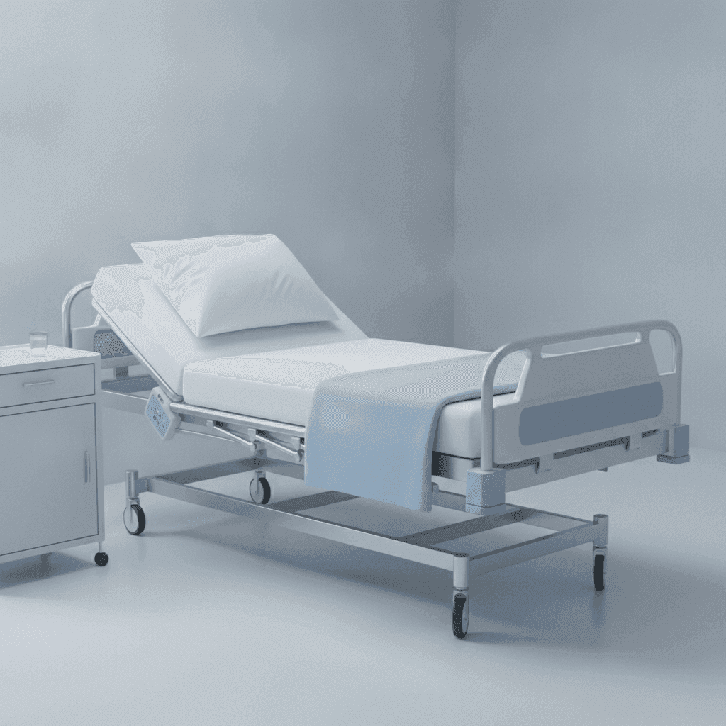
The skull is a closed box. Pressure falls when venous blood can leave the head and when you avoid spikes in cerebral blood volume or intrathoracic/intra-abdominal pressure. Head of bed about 30° with the neck midline keeps jugular veins open; a tight collar, extreme hip flexion, or lying flat for long stretches dams venous return and ICP climbs.
| Do | Avoid |
|---|---|
| HOB ~30°, neck midline, loosen collar if it compresses veins | Flat for long periods; extreme hip flexion |
| Calm environment; space care; stool softeners to limit Valsalva | Clustering suction, turns, and painful procedures |
| Brief, necessary suction with pre-oxygenation | Prolonged suction or coughing fits you can prevent |
| Maintain ordered CO₂ / ventilation goals | Hypercapnia and hypoxia (both raise cerebral blood volume) |
Why space care and soften stool: each suction pass, turn, and painful stimulus can transiently raise ICP. Stack them and you create one prolonged spike the brain cannot clear. Valsalva from constipation does the same through venous backup. Brief suction with pre-oxygenation clears the airway without trading hypoxia or a coughing fit for a pressure surge. Ordered ventilation goals matter because hypercapnia and hypoxia both dilate cerebral vessels and raise blood volume inside the skull. The trap is chasing comfort positioning or “getting everything done at once” while CO₂ and O₂ drift.
The distractor that looks caring is laying the client flat to rest, clustering AM care for efficiency, or suctioning until the airway sounds perfect. Flat and clustered care raise pressure; prolonged suction costs more ICP than a short, oxygenated pass. Edge case: spinal precautions or an ordered flat position after certain injuries override the usual 30° rule: follow the stem’s clearance and still keep the neck neutral when you can.
Safety
A sudden drop in LOC, a new blown pupil, or posturing is not a “reassess in an hour” finding. Notify the provider and protect ABCs.
Head injury specifics
Head trauma adds fracture paths, CSF leaks, and seizure risk on top of the ICP rules above. Basilar skull fracture signs (raccoon eyes, Battle sign, CSF from nose or ear) mean the base of the skull may be open. A nasal NG tube can track into the cranial vault, so do not place one through the nose unless imaging and the provider have cleared that route.
- Basilar skull fracture cues: raccoon eyes, Battle sign, CSF otorrhea/rhinorrhea: do not insert NG tubes through the nose unless cleared.
- CSF leak: allow drainage to a pad as ordered; do not pack the nose/ear tightly or teach nose-blowing.
- Seizure precautions when the cortex is irritated; have suction and oxygen ready.
- Neuro checks on a schedule: compare to the client’s baseline, not a generic “normal.”
CSF leaking from nose or ear is a pathway for meningitis, not a mess to pack shut. Allow drainage to a pad as ordered, skip nose-blowing and tight packing, and watch for fever, stiff neck, and worsening headache. Cortical irritation after injury can trigger seizures: precautions and ready suction/oxygen protect the airway when a fit hits. Serial neuro checks only work if you compare to *this* client’s baseline; a GCS that looks “almost normal” can still be a drop from their earlier exam.
The distractor that looks decisive is packing rhinorrhea, teaching the client to blow hard to clear the nose, or delaying escalation until pupils match a textbook late picture. Packing and Valsalva worsen leak risk and pressure dynamics; waiting for a perfect Cushing triad misses the window. Swallow safety and full stroke-style neuro assessment live in Stroke and Neuro Assessment. Use that chapter when the stem turns on dysphagia or a focused neuro exam rather than ICP mechanics.
Epidural vs subdural patterns
Epidural hematoma is often arterial (classic middle meningeal pattern) after trauma: loss of consciousness, a lucid interval, then rapid decline. That lucid gap is not clearance to send the client home without instructions. Subdural hematoma is often venous bridging-vein bleeding. It may evolve slower, especially in older adults or clients on anticoagulants, with progressive confusion days after a fall. Both need serial neuro checks and escalation when LOC slips. Concussion teaching still includes waking parameters and return precautions as ordered.
| Bleed pattern | Exam fingerprint |
|---|---|
| Epidural | Trauma + lucid interval + sudden decline |
| Acute subdural | Trauma + progressive LOC change |
| Chronic subdural (elder) | Fall days ago + new confusion; anticoagulants raise risk |
Osmotherapy (mannitol pattern)
Hyperosmolar agents pull water from brain tissue into the vascular space. Expect heavy urine output. Watch fluid balance, sodium, and neuro response. Use a filter as ordered for mannitol crystals; hold and clarify if the solution is crystallized. The exam wants recognition that this is acute ICP therapy, not a casual diuretic teaching moment. Hypertonic saline appears on some protocols with the same close sodium and neuro monitoring.
How to reason under time pressure
- Is the airway and oxygenation intact?
- Is LOC changing from this client’s baseline?
- Fix positioning and stop ICP-raising actions before chasing every lab.
- Escalate late signs immediately. Do not wait for a perfect triad.
Revision
Must know
- 1Early ICP rise: change in LOC is the most sensitive cue. Late: Cushing triad (hypertension with widening pulse pressure, bradycardia, irregular respirations).
- 2Head of bed about 30°, head midline, avoid hip flexion and clustering care that spikes ICP.
- 3Do not suction longer than needed; pre-oxygenate; avoid Valsalva, tight collars, and flat prolonged positioning when ICP is high.
- 4CSF leak from nose/ear: do not pack aggressively or blow the nose; report; watch for meningitis signs.
- 5Mannitol / hyperosmolar therapy: expect diuresis; watch output, electrolytes, and neuro status.
- 6Unequal or fixed pupils and posturing are emergency neuro deterioration: notify provider now.
Memory hooks
LOC first, Cushing late
Altered consciousness shows up before the classic vital-sign triad. Do not wait for Cushing findings to act on rising ICP.
Midline and thirty
Neutral neck and roughly 30° head elevation promote venous drainage from the skull.
On the exam
How it's tested
Stems show a head-injury client with restlessness, pupil change, or a nursing action that raises ICP. Distractors lay the client flat, cluster painful care, pack a CSF rhinorrhea, or treat Cushing triad as early and reassuring.
More in adult health
All topics- Acute coronary syndromesNCLEX ACS chapter: STEMI reperfusion, nitroglycerin and cath-site priorities, cardiogenic shock, tamponade and endocarditis cues, and valvular stenosis vs regurgitation.Read
- Acute respiratory emergenciesNCLEX airway map: failure cues, PE vs pneumothorax vs obstruction, oxygen and positioning priorities, and when to escalate.Read
- AnemiasNCLEX hematology chapter: iron, B12, and folate deficiency teaching, anemia activity tolerance, and sickle-cell crisis priorities with emergency red flags.Read
- Antimicrobial therapyNCLEX antimicrobial chapter: allergy checks, finishing the course, vancomycin flushing, aminoglycoside toxicity, TB drug teaching, and common topical agents.Read